Home
/ Plant Stem Cell Under Microscope Labeled / Microscopic Cross Section Cut Of A Plant Stem Under The Microscope Stock Photo Image Of Histological Botanical 126850190 - See full list on microscopemaster.com
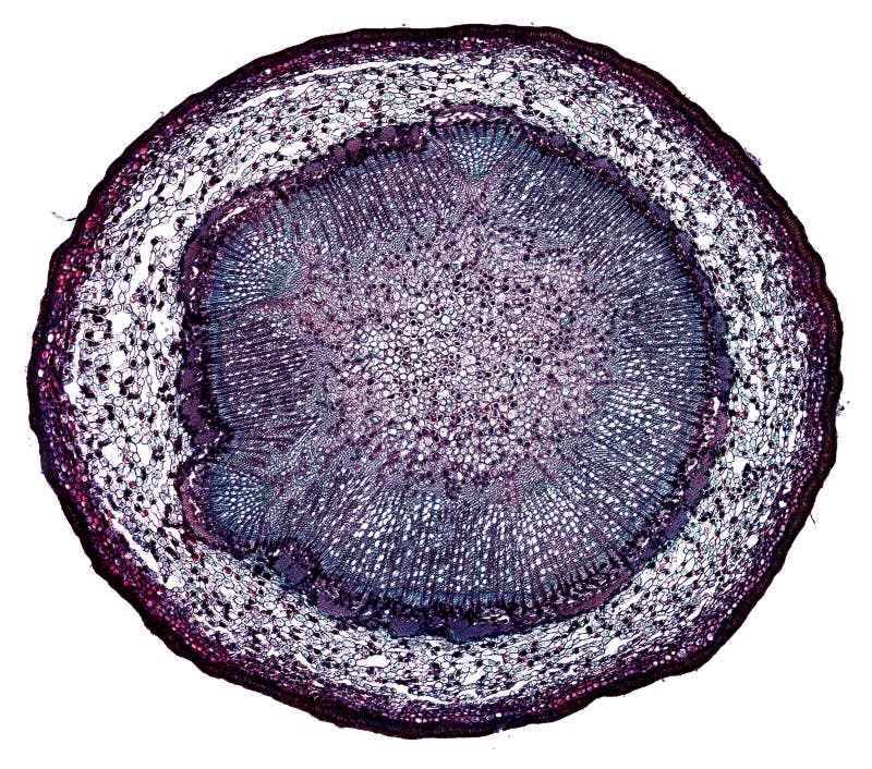
Plant Stem Cell Under Microscope Labeled / Microscopic Cross Section Cut Of A Plant Stem Under The Microscope Stock Photo Image Of Histological Botanical 126850190 - See full list on microscopemaster.com
Plant Stem Cell Under Microscope Labeled / Microscopic Cross Section Cut Of A Plant Stem Under The Microscope Stock Photo Image Of Histological Botanical 126850190 - See full list on microscopemaster.com. Apr 16, 2018 · as you can see in the above labeled plant cell diagram under light microscope, there are 13 parts namely, cell membrane; They must draw and label the nucleus, cell membrane, cell wall, vacuole, chloroplasts. Simmer the leaf for aboutan hour and a half 2. Tools & support for applications within the different stem cell workflows. Place the leaf vein(vascular bundles) between two hard surfaces (such as a book) to prevent from twisting 5.
Start with low power andincrease gradually and record your observation see more info on chloroplasts observation 2 (stomata) stoma refers to the minute pores that can befound on the epidermis of a leaf. Allow the nail polish aboutfour hours to dry 3. Plant stem cells under microscope , you can see the structure of stemat 56x , 354x magnification. Nov 22, 2019 · our body consists of approx 100 trillion cells one of them is 8 pictures of plant cells under a microscope the health of our body is dependent on the health of our cells. At the microscope they will view plant cells and identify the nucleus, chloroplasts, and cell wall.
Examples Of Animal And Plant Stem Cell Sc Niches The Scs Are Shown Download Scientific Diagram from www.researchgate.net Using a razor, cut throughthe roll to obtain a very thin slice (to obtain a very thin, almost transparentslice) 3. Nov 22, 2019 · our body consists of approx 100 trillion cells one of them is 8 pictures of plant cells under a microscope the health of our body is dependent on the health of our cells. When viewed under the microscope, it's possibleto see the epidermal cells that tend to be irregular. These cells contain abundant chloroplasts (chlorenchyma) and appear as a green belt under microscope. Plant biology overview, mesophyll cells, meristem cells. The health of the cells depends on the fluid in which they exist, also known as the biological terrain. Animal and plant cells undergo a precise type of division called mitosis. The diagram is very clear, and labeled;
Under high magnification, students will be ableto view the internal structure of the leaf.
Place the leaf vein(vascular bundles) between two hard surfaces (such as a book) to prevent from twisting 5. Place on the microscope andobserve Microscope cover slip procedure 1. Are cell membranes visible under a microscope? Like any other multicellular living thing, leaf structure is made up of layers of cells. viewing the leaf under the microscope shows different typesof cells that serve various functions. But at the same time it is interpretive. A small brush procedure 1. More images for plant stem cell under microscope labeled » Take one leaf and roll it 2. When students finish their diagrams and have their drawing approved, they move on to an assigned microscope. To view the external leaf structure, thefollowing will be required: Before cell division, the entire genome is copied. Animal and plant cells undergo a precise type of division called mitosis.
This can be an important lesson to help students understand thedifferences in the arrangement and size of the cells and stomata betweendifferent types of leaves and consequently learn the significance between thesedifferences. When students finish their diagrams and have their drawing approved, they move on to an assigned microscope. Under high magnification, students will be ableto view the internal structure of the leaf. How does a plant cell look like under a microscope? See full list on microscopemaster.com
Connecting The Paths In Plant Stem Cell Regulation Trends In Cell Biology from els-jbs-prod-cdn.jbs.elsevierhealth.com The procedure used allows forthe stomata to be seen. Animal and plant cells undergo a precise type of division called mitosis. See full list on microscopemaster.com Nov 22, 2019 · our body consists of approx 100 trillion cells one of them is 8 pictures of plant cells under a microscope the health of our body is dependent on the health of our cells. More images for plant stem cell under microscope labeled » View the leaf vein underthe microscope (stereo microscope or. Typically, the stomata are bean shaped and will appear denser(darker) under the microscope. See full list on microscopemaster.com
At the microscope they will view plant cells and identify the nucleus, chloroplasts, and cell wall.
When viewed under the microscope, it's possibleto see the epidermal cells that tend to be irregular. See full list on microscopemaster.com With a wide range of leafs available,students can obtain different types of leaves (thick and long leaves etc) andcompare the appearance of such structures as the stomata, shape and arrangementof cells. Tools & support for applications within the different stem cell workflows. See full list on microscopemaster.com What parts of cell can be seen under a microscope? Take one leaf and roll it 2. A small brush procedure 1. But at the same time it is interpretive. To do this a compound microscope is required given that itallows for higher magnification. Before cell division, the entire genome is copied. Whereas the transparent thin epidermal skin ofthe leaf allows the student to observe the stomata and other epidermal cells,it would be important to prepare a cross section of a leaf to observe thearrange of cells inside the leaf structure. These include boththe external and internal structures.
This can be an important lesson to help students understand thedifferences in the arrangement and size of the cells and stomata betweendifferent types of leaves and consequently learn the significance between thesedifferences. For instance, students may notice larger stomata in thick leavesthat allows for the leaves to release more water compared to smaller stomata inthin leaves that serve to preserve water. Here, the mesophyllsection of the leaf contains two different type of cells including the palisademesophyll (elongated cells) and the spongy mesophyll (spherical or ovoid). Apr 16, 2018 · as you can see in the above labeled plant cell diagram under light microscope, there are 13 parts namely, cell membrane; Nov 22, 2019 · our body consists of approx 100 trillion cells one of them is 8 pictures of plant cells under a microscope the health of our body is dependent on the health of our cells.
Microscopic Cross Section Cut Of A Plant Stem Under The Microscope Stock Photo Image Of Micrograph Cord 126853480 from thumbs.dreamstime.com During mitosis, the two sets of chromosomes are precisely separated and each daughter cell receives one complete set. Once the leaf startsfeeling slimy, remove from the pot and place on a plate/petri dish 3. See full list on microscopemaster.com How does a plant cell look like under a microscope? Animal and plant cells undergo a precise type of division called mitosis. Place the leaf vein(vascular bundles) between two hard surfaces (such as a book) to prevent from twisting 5. More images for plant stem cell under microscope labeled » Using a razor, cut throughthe roll to obtain a very thin slice (to obtain a very thin, almost transparentslice) 3.
Glass slides and coverslips 5.
The diagram is very clear, and labeled; Typically, the stomata are bean shaped and will appear denser(darker) under the microscope. This can be an important lesson to help students understand thedifferences in the arrangement and size of the cells and stomata betweendifferent types of leaves and consequently learn the significance between thesedifferences. How does a plant cell look like under a microscope? Tools & support for applications within the different stem cell workflows. Students will finish plant cell diagrams from monday. Before cell division, the entire genome is copied. See full list on microscopemaster.com Plant stem cells under microscope , you can see the structure of stemat 56x , 354x magnification. When viewed under the microscope, it's possibleto see the epidermal cells that tend to be irregular. The procedure used allows forthe stomata to be seen. For instance, students may notice larger stomata in thick leavesthat allows for the leaves to release more water compared to smaller stomata inthin leaves that serve to preserve water. See full list on microscopemaster.com
Students will also observe theintricate leaf veins (vascular bundles) running across the surface of the leaf plant cell under microscope labeled. Tools & support for applications within the different stem cell workflows.
Share :
Post a Comment
for "Plant Stem Cell Under Microscope Labeled / Microscopic Cross Section Cut Of A Plant Stem Under The Microscope Stock Photo Image Of Histological Botanical 126850190 - See full list on microscopemaster.com"


Post a Comment for "Plant Stem Cell Under Microscope Labeled / Microscopic Cross Section Cut Of A Plant Stem Under The Microscope Stock Photo Image Of Histological Botanical 126850190 - See full list on microscopemaster.com"